


The Imaging and Phenotyping Core of the Lady Davis Institute for Medical Research (Animal)
Name of entity
The Imaging and Phenotyping Core of the Lady Davis Institute for Medical Research
Entity type
Plateform
Keywords
Imaging, ultrasound, bioluminescence, fluorescence, cardiovascular, echocardiography, electrocardiography, catheterization, hematology sphygmomanometer, telemetry, blood pressure, vascular remodeling, vascular stiffness, oncology, tumors, metastasis, Ami HT, VETSCAN® HM5, VEVO 3100
Area of expertise
Study of cardiovascular function and oncological imaging in vivo
Services offered
Our service is based on the following equipment:
– A bioluminescence or fluorescence imaging system (Ami HT)
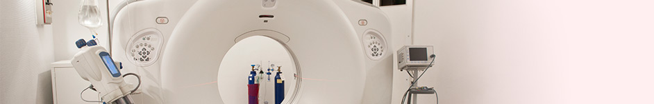
– A high frequency ultrasound system (VEVO 3100)
– An EMKA physiological parameter acquisition and analysis system including pressure, velocity and surface ECG
– Millar micro catheters
– A caudal plethysmography of Hatteras
– A telemetry system from Data Science International
– A veterinary hematology analyzer (VETSCAN® HM5)
More specifically, we offer:
Study of the cardiovascular system: echocardiography, surface electrocardiography, blood pressure by sphygmomanometer, Millar catheter or telemetry, measurement of the intima media and vascular stiffness (pulse wave velocity) by ultrasound.
Oncological imaging study in vivo: Measurement of the size in two or three dimensions and angiogenesis of primary tumors or metastases by ultrasound, monitoring of tumors and development of metastases by bioluminescence or fluorescence imaging of cancer cells in vivo.
Véronique Michaud
ldi_phenotyping@ladydavis.ca
https://www.ladydavis.ca/fr/a-propos/plateformes-de-recherche/imagerie-et-phenotypage/
Address
Lady Davis Institute for Medical Research
3999 Ch. Côte Ste-Catherine,
Montréal, Québec,
H3T 1E2